Gram Positive Flow Chart
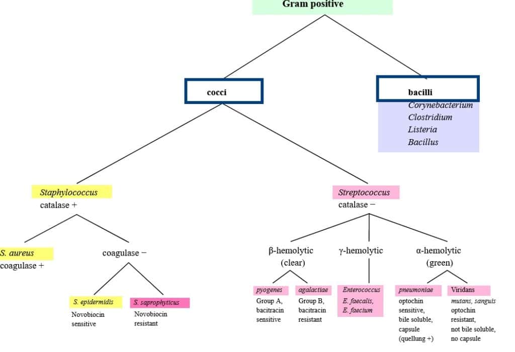
Gram Positive Flow Chart - Coagulase negative staphylococci in multiple blood cultures. Lugdunensis found in abscesses and serious wounds; Give an example of a bacterium of high g+c and low g+c group commonly associated with each category. Salt tolerance test hemolysis test bacitracin test. Web gram staining is the common, important, and most used differential staining techniques in microbiology, which was introduced by danish bacteriologist hans christian gram in 1884. The bacterial cell wall of these organisms have thick peptidoglycan layers, which take up the purple/violet stain. During the gram staining process — a test that experts use to view the bacteria under a microscope — they appear purple or. Web flow chart of gram positive organisms for infectious diseases unknown labs at kcom. Gram positive (g+), catalase positive (+) cocci. Web gram positive cocci obligate anaerobic peptostreptococcus spp., peptinophilus spp., parvimonas spp., anaerococcus spp., atopobium spp., f. Web the following figures illustrate decision algorithms to facilitate bacterial identification. Learn vocabulary, terms, and more with flashcards, games, and other study tools. The bacterial cell wall of these organisms have thick peptidoglycan layers, which take up the purple/violet stain. Web the six flow charts we’ll be discussing are: During the gram staining process — a test that experts use to view the bacteria under a microscope — they appear purple or. Web gram staining is the common, important, and most used differential staining techniques in microbiology, which was introduced by danish bacteriologist hans christian gram in 1884. Gram positive (g+), catalase positive (+) cocci. Web the simulator features an interactive flow chart in which users can learn more about the biochemical tests and follow pathways to specific organisms. Coagulase negative staphylococci in multiple blood cultures. Identify similarities and differences between high g+c and low g+c bacterial groups. Interpret the results of biochemical methods. Web gram staining is the common, important, and most used differential staining techniques in microbiology, which was introduced by danish bacteriologist hans christian gram in 1884. Flow chart of gram positive organisms created date: Web aerobic gram positive rods flowchart. Web the six flow charts we’ll be discussing are: Web flow chart of gram positive organisms for infectious diseases unknown labs at kcom. Coagulase negative staphylococci in multiple blood cultures. Web aerobic gram positive rods flowchart. Web the following figures illustrate decision algorithms to facilitate bacterial identification. Web the simulator features an interactive flow chart in which users can learn more about the biochemical tests and follow pathways to. Gram positive (g+), catalase positive (+) cocci. Flow chart of gram positive organisms created date: Web the simulator features an interactive flow chart in which users can learn more about the biochemical tests and follow pathways to specific organisms. This test differentiate the bacteria into gram positive and gram negative bacteria, which helps in the classification and differentiations of microorganisms.. Flow chart of gram positive organisms created date: Web start studying gram positive bacteria flow chart. Associate various biochemical tests with their correct applications. Web the six flow charts we’ll be discussing are: Web use flowcharts and identification charts to identify some common aerobic gram positive microorganisms. Web aerobic gram positive cocci flowchart. Gram positive (g+), catalase positive (+) cocci. Web tmcc microbiology resource center unknown identification work flow flowchart. Associate various biochemical tests with their correct applications. Interpret the results of biochemical methods. Web the six flow charts we’ll be discussing are: Web gram positive bacteria types and classification. The bacterial cell wall of these organisms have thick peptidoglycan layers, which take up the purple/violet stain. Web aerobic gram positive cocci flowchart. Users can click on the test name to learn more about that test as well as see images or videos presenting. Flow chart of gram positive organisms created date: Web tmcc microbiology resource center unknown identification work flow flowchart. Web gram positive bacteria types and classification. Web aerobic gram positive rods flowchart. Web flow chart of gram positive organisms for infectious diseases unknown labs at kcom. Web the following figures illustrate decision algorithms to facilitate bacterial identification. Coagulase negative staphylococci in multiple blood cultures. Coagulase test hemolysis test novobiocin test. Web the simulator features an interactive flow chart in which users can learn more about the biochemical tests and follow pathways to specific organisms. Flow chart of gram positive organisms created date: Web gram positive cocci obligate anaerobic peptostreptococcus spp., peptinophilus spp., parvimonas spp., anaerococcus spp., atopobium spp., f. Web tmcc microbiology resource center unknown identification work flow flowchart. Salt tolerance test hemolysis test bacitracin test. Web gram staining is the common, important, and most used differential staining techniques in microbiology, which was introduced by danish bacteriologist hans christian gram in 1884.. Associate various biochemical tests with their correct applications. Lugdunensis found in abscesses and serious wounds; Web flow chart of gram positive organisms for infectious diseases unknown labs at kcom. Web gram staining is the common, important, and most used differential staining techniques in microbiology, which was introduced by danish bacteriologist hans christian gram in 1884. Web the six flow charts. Lugdunensis found in abscesses and serious wounds; During the gram staining process — a test that experts use to view the bacteria under a microscope — they appear purple or. Web tmcc microbiology resource center unknown identification work flow flowchart. Flow chart of gram positive organisms created date: Learn vocabulary, terms, and more with flashcards, games, and other study tools. Interpret the results of biochemical methods. Web gram positive bacteria types and classification. Coagulase negative staphylococci in multiple blood cultures. Identify similarities and differences between high g+c and low g+c bacterial groups. Web the simulator features an interactive flow chart in which users can learn more about the biochemical tests and follow pathways to specific organisms. Web gram positive cocci obligate anaerobic peptostreptococcus spp., peptinophilus spp., parvimonas spp., anaerococcus spp., atopobium spp., f. Associate various biochemical tests with their correct applications. Web the six flow charts we’ll be discussing are: Web use flowcharts and identification charts to identify some common aerobic gram positive microorganisms. Web start studying gram positive bacteria flow chart. Web gram staining is the common, important, and most used differential staining techniques in microbiology, which was introduced by danish bacteriologist hans christian gram in 1884.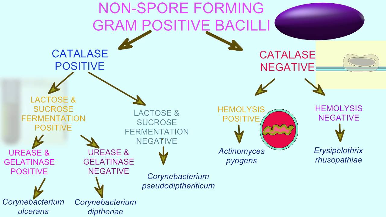
Gram Positive Flow Chart
Gram positive flow chart BS Nursing BatStateU Studocu
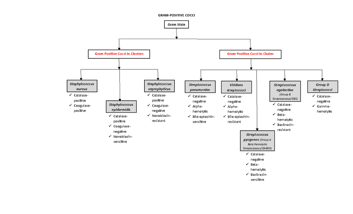
Gram Positive Cocci Flow Chart Streptococcus Prokaryote

Gram Positive Bacteria Overview Identification Algorithm
GramPositive Bacteria Characteristics, List, Cell wall composition

Gram Positive Flow Chart 1 Diagram Quizlet

Gram Positive Bacteria Flow Chart Diagram Quizlet

Flow Chart For Gram Positive Cocci

Gram Positive Bacteria Flow Chart

Gram Positive Bacteria Flow Chart Diagram Quizlet
Users Can Click On The Test Name To Learn More About That Test As Well As See Images Or Videos Presenting Positive And Negative Reactions.
Coagulase Test Hemolysis Test Novobiocin Test.
Give An Example Of A Bacterium Of High G+C And Low G+C Group Commonly Associated With Each Category.
Web Aerobic Gram Positive Cocci Flowchart.
Related Post:
