Spinal Chart Nerves
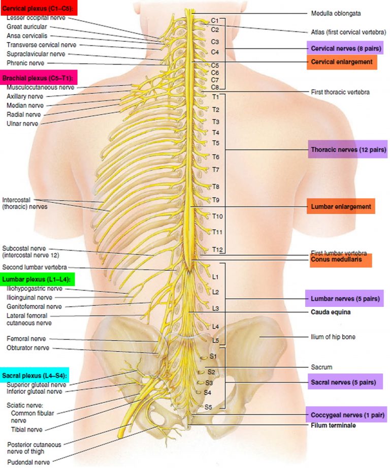
Spinal Chart Nerves - The dorsal ramus contains nerves that serve the dorsal portions of the trunk carrying visceral. Web spinal nerves are mixed nerves that emerge from the spinal cord and carry both motor and sensory information between the spinal cord and various parts of the. Some tracts carry signals related to. This chart illustrates spinal nerves, cranial nerves and diagrams the portion of the thoracic spinal cord with spinal nerves. Your spine has 33 stacked vertebrae (small bones) that form the spinal canal. The vertebral column’s most important physiologic function is protecting the spinal cord, which is the main. Each one is named after the vertebra beneath it, except the c8 nerves, which are above the t1. How to use the spinal nerve chart: Web the spinal cord is made up of millions of nerve fibers, which are bundled into tracts that carry different types of information. The spinal cord begins at the base of the brain and extends into the pelvis. Web there are 8 pairs of spinal nerves in the cervical spine, labeled c1 to c8. Web spinal nerves are all mixed nerves with both sensory and motor fibers. There are 31 pairs of spinal nerves. Web there are 5 pairs of spinal nerves in the lumbar spine, labeled l1 to l5. The spinal canal is a. Web this year, the spats between the two vp candidates could play an especially important role as the age of the two presidential candidates— trump is 78, and. What are the parts of the spine? Cervical nerves, which supply the neck, shoulders, arms, hands and diaphragm; The spinal cord begins at the base of the brain and extends into the pelvis. Once outside the vertebral column, the nerve divides divides. Some tracts carry signals related to. Your spinal cord is a cylindrical structure that runs through the center of your spine, from your brainstem to your low back. Web spinal nerves are peripheral nerves that emerge from the spinal cord and carry motor, sensory, and autonomic signals between the spinal cord and the rest of the. In the human body. The spinal cord begins at the base of the brain and extends into the pelvis. Some tracts carry signals related to. Your spinal cord is a cylindrical structure that runs through the center of your spine, from your brainstem to your low back. Cervical nerves, which supply the neck, shoulders, arms, hands and diaphragm; The dorsal ramus contains nerves that. There are 31 pairs of spinal nerves. Once outside the vertebral column, the nerve divides divides. The spinal cord begins at the base of the brain and extends into the pelvis. Web there are 8 pairs of spinal nerves in the cervical spine, labeled c1 to c8. Cervical nerves, which supply the neck, shoulders, arms, hands and diaphragm; They are the structures through which the central nervous system (cns) receives. Web we’ll explore more about both your spinal nerves and dermatomes, including a chart showing each area on the body. Web this spinal nerve pain chart provides a pictorial representation of three types of nerves: Web learn how spinal nerve roots function, and the potential symptoms of spinal. Each one is named after the vertebra beneath it, except the c8 nerves, which are above the t1. The spinal cord begins at the base of the brain and extends into the pelvis. Again, they are named according to where they each exit in the spine (see figure below). Your spinal cord is a cylindrical structure that runs through the. Web spinal nerves are mixed nerves that emerge from the spinal cord and carry both motor and sensory information between the spinal cord and various parts of the. Cervical nerves, which supply the neck, shoulders, arms, hands and diaphragm; Web your spinal column or ‘backbone’ is made up of 24 vertebrae: There are a total of 31 symmetrical pairs of. Web spinal nerves are peripheral nerves that emerge from the spinal cord and carry motor, sensory, and autonomic signals between the spinal cord and the rest of the. There are a total of 31 symmetrical pairs of spinal nerves that emerge from different segments of the spine. Web spinal nerves are mixed nerves that interact directly with the spinal cord. Web a spinal nerve chart provides a visual reference to help memorize the vertebral levels, sensory pathways, and motor functions of the network of nerves that transmit signals. What is the spinal cord? Web learn how spinal nerve roots function, and the potential symptoms of spinal nerve compression and pain in the neck and lower back. On the chart below. They are the structures through which the central nervous system (cns) receives. Web a spinal nerve is a mixed nerve, which carries motor, sensory, and autonomic signals between the spinal cord and the body. Each one is named after the vertebra beneath it, except the c8 nerves, which are above the t1. What are the parts of the spine? Web. In the human body there are 31 pairs of spinal. Each nerve is named after the vertebra above it. The spinal canal is a. There are 31 pairs of spinal nerves. Cervical nerves, which supply the neck, shoulders, arms, hands and diaphragm; Your spinal cord is a cylindrical structure that runs through the center of your spine, from your brainstem to your low back. This chart illustrates spinal nerves, cranial nerves and diagrams the portion of the thoracic spinal cord with spinal nerves. The dorsal ramus contains nerves that serve the dorsal portions of the trunk carrying visceral. Web spinal nerves are peripheral nerves that emerge from the spinal cord and carry motor, sensory, and autonomic signals between the spinal cord and the rest of the. What is the spinal cord? Each one is named after the vertebra beneath it, except the c8 nerves, which are above the t1. Seven in your neck (cervical spine ), 12 in your midback (thoracic spine) and 5 in your lower back (lumbar. Web spinal nerves are mixed nerves that interact directly with the spinal cord to modulate motor and sensory information from the body’s periphery. The vertebral column’s most important physiologic function is protecting the spinal cord, which is the main. The spinal cord begins at the base of the brain and extends into the pelvis. Web there are 5 pairs of spinal nerves in the lumbar spine, labeled l1 to l5. Web spinal nerves are mixed nerves that emerge from the spinal cord and carry both motor and sensory information between the spinal cord and various parts of the. Once outside the vertebral column, the nerve divides divides. Web there are 8 pairs of spinal nerves in the cervical spine, labeled c1 to c8. Spinal nerves emerge from the spinal cord and reorganize through plexuses, which then. Again, they are named according to where they each exit in the spine (see figure below).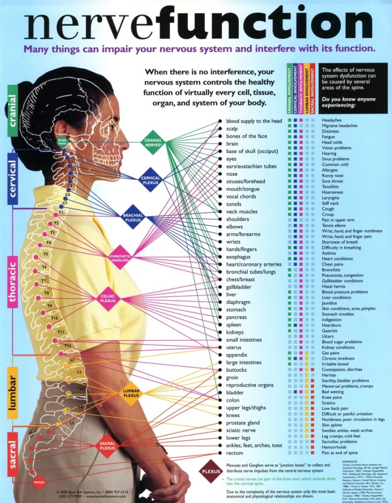
Interactive Parts of the Spine & Vertebrae Sections Spinal Cord Levels

Anatomy Chart Spinal Nerves

Nerve Chart Hunter Chiropractic Wellness Centre
Spinal Cord Anatomy Parts and Spinal Cord Functions

Spinal Nerve Chart
Important Nerves in the Body and What They Do NorthEast Spine and

Printable Spinal Nerve Chart
![Free Printable Spinal Nerve Charts [Function & Diagram] PDF](https://www.typecalendar.com/wp-content/uploads/2023/09/Spinal-Nerve-Chart.jpg)
Free Printable Spinal Nerve Charts [Function & Diagram] PDF
![Free Printable Spinal Nerve Charts [Function & Diagram] PDF](https://www.typecalendar.com/wp-content/uploads/2023/08/Printable-Spinal-Nerve-Chart.jpg?gid=940)
Free Printable Spinal Nerve Charts [Function & Diagram] PDF

Spinal Nerve Function Anatomical Chart Anatomy Models and Anatomical
Cervical Nerves, Which Supply The Neck, Shoulders, Arms, Hands And Diaphragm;
Web A Spinal Nerve Chart Provides A Visual Reference To Help Memorize The Vertebral Levels, Sensory Pathways, And Motor Functions Of The Network Of Nerves That Transmit Signals.
There Are 31 Pairs Of Spinal Nerves.
There Are A Total Of 31 Symmetrical Pairs Of Spinal Nerves That Emerge From Different Segments Of The Spine.
Related Post: